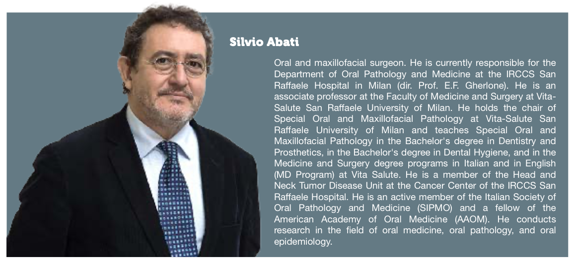
San Raffaele's Dr Silvio Abati: A few minutes to save a life

Interview by Paola Brambilla with professor Silvio Abati, responsible for the Departmental Center of Pathology and Oral Medicine of the Department of Dentistry at the IRCCS Ospedale San Raffaele in Milan. Published in Il dentista moderno, June 2024, pp. 16-18. Featuring Studium Genetics SG-OCRA test.

Download the full article (Italian).
[Unofficial, automatic translation]
During their professional life, every dentist encounters on average three to five malignant tumors of the mouth, often unfortunately in advanced stages. This condition could be prevented by investing a few minutes during routine visits or oral hygiene sessions to perform an accurate visual examination of the oral cavity of their patients: a simple gesture, but very useful for detecting potential pre-tumoral and precancerous lesions, which, if recognized and treated in time, would not transform into tumors.
Despite its crucial importance, the early diagnosis of malignant tumors of the mouth still represents a complex area of prevention and is one of the most serious problems for the role of the dentist in the context of diseases not strictly related to teeth and gums. We discussed this with Professor Silvio Abati, responsible for the Departmental Center of Pathology and Oral Medicine at the Dental Clinic-Department of Dentistry of the IRCCS Ospedale San Raffaele in Milan, who dedicates his studies and research to the prevention and care of oral diseases.
Professor Abati, how widespread are mouth tumors today?
The statistics on mouth tumors that concern dentists refer to those of the visible part of the mouth, while many statistics include not only the oral cavity but also the oropharynx. In Italy, there are approximately 6,000 to 8,000 cases of mouth tumors per year. In the past, oral cavity tumors were mainly found in people over 65 years old, but today they are very common in the 50-year-old age group, and many cases are also found in patients between 30 and 35 years old. 50% of malignant tumors are made up of over 90% squamous cell carcinomas of the epithelium and occur more easily on the tongue, especially on the ventral surface or edges. In their professional life, a dentist may see, on average, three to five malignant tumors of the mouth, but they also have the opportunity to discover, during routine visits, pre-tumoral or precancerous lesions, which should be recognized and treated immediately.
What can the dentist do concretely?
Their role is important for intercepting suspicious lesions. It is necessary for the dentist to systematically visit the oral mucosa of their patients at least twice a year, performing an accurate examination of the oral cavity that includes inspection and palpation of all oral mucosa sites. Particular attention should be paid to all variations from the norm of tissue characteristics, considering the surface features, color, consistency, and mobility. This is especially important when visiting patients with a smoking or drinking habit, which represent significant risk factors, especially if associated.
What signs should raise suspicions?
The manifestations that should raise suspicions and suggest the possibility of an oral tumor include localized and persistent changes in the color of the mucosa, such as white, red, or mixed white and red patches; ulcers that do not heal or bleed easily; tumors, thickening, or productive lesions that cause tissue hardening. In more advanced stages, oral cancer manifests with nodular, ulcerated, vegetating, or infiltrating lesions; sometimes the patient complains of pain and difficulty with normal swallowing and mastication or notices unusual changes in phonation.
How important is the time factor in diagnosis?
The time factor is truly crucial. This is confirmed by the fact that, due to the Covid-related shutdown, tumor diagnoses have increased. Unfortunately, specialized centers still receive patients with advanced tumors, forced to undergo significant compromise of their quality of life. The difference between a good prognosis, which requires limited resources for therapy, and a poor prognosis, which involves enormous costs, is sometimes determined solely by the ability to make a correct diagnosis one year earlier or later.
How do these preventive tools work?
The clinical use of autofluorescence is based on the evidence that both premalignant lesions, such as epithelial dysplasia, and cancer cause visible changes in the autofluorescence of the oral mucosa, making it possible to visualize potentially cancerous or frankly cancerous lesions better and with greater ease, as well as select with greater precision the tissue portions to be submitted to biopsy. The epigenetic test, which is performed through genetic analysis of cells collected from the oral mucosa with a brush, allows for a non-invasive first investigation in identified sites. Our center was the first in Italy to systematically use the Ocra salivary test developed by Studium Genetics, a spin-off of Alma Mater Studiorum - University of Bologna. The test has proven to be a particularly useful tool for analyzing suspicious lesions in patients over 40 who smoke or drink heavily, who are more exposed to the risk of developing oral cavity tumors.
What are the costs necessary to acquire these preventive tools?
The special glasses or other devices for visualizing autofluorescence and the epigenetic test on superficial cells have limited and more than accessible costs when compared to the enormous advantage they can offer.
Why is it so important to intervene early?
Early diagnosis of the patient, when the tumor is less than two centimeters, increases the five-year survival rate to 90%. When, instead, the tumor is detected at the third or fourth stage, with dimensions greater than four centimeters and having already involved lymph nodes, the five-year survival rate drops to below 20-30%. The surgical approach also differs. Removing a small tumor of five millimeters under local anesthesia is very different from removing, for example, a five-centimeter tumor of the tongue that has already invaded surrounding anatomical structures: it requires an expert surgical team and an intervention that may last 10-12 hours. The interventions often result in highly mutilating consequences, even when reconstructions of the affected areas are performed. Furthermore, it is necessary to refer to structures capable of handling this type of surgery. Additionally, the healthcare costs for treating a small tumor are greatly reduced, while the necessary expenses for a major intervention and subsequent follow-up represent significant figures.
How much time passes, on average, from diagnosis to recovery?
In the most serious cases, the recovery times, which involve radiation therapy and chemotherapy, are very long: it takes over three or four months, not counting that the patient's residual quality of life is often severely compromised, especially when significant demolitions of mouth and face structures are necessary. The pre-intervention diagnostic phase, necessary for planning surgical and radiation therapy, requires the results investigations such as biopsy, magnetic resonance imaging with contrast medium, CT, PET, and all that, in some areas of Italy, require rather long times.
How are pre-tumoral lesions treated, instead?
